Types of vision tests: what do they measure?
Whether you have healthy or poor vision, it is important to see an eye doctor regularly for proper testing. The overall health of your vision will help your doctor determine when further intervention is necessary.
There are certain types of tests that are performed for everyone during a comprehensive eye examination, such as visual acuity testing. For some people with specific vision issues, additional testing may be performed, such as testing for color blindness. The doctor will begin by evaluating the state of your vision and eye health. From here, they can determine which tests are appropriate for your comprehensive eye examination.
The following types of tests highlight the wide variety of vision tests that are available.
Visual Acuity Testing
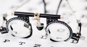
This type of test is done to determine how sharp your vision is. The doctor will usually have you look at an eye chart for this examination. They will ask you to read the numbers or letters that are present on a specific line.
In some cases, doctors give you a handheld chart. This one is used to determine how sharp your vision is close up.
Color Blindness Testing
The Ishihara Color Vision Test is the color blindness test that is the most widely used.
For this test, you are asked to look at a booklet. There will be dots of varying brightness, colors, and sizes. If you have normal color vision, you will be able to pick out the number that is present among the dots.
Ocular Motility Testing
This test is done to see if your eyes are moving as they should. It tests your ability to accurately focus on two separate objects and follow a moving object.
This test is important because if you have abnormal eye movements, you are at a higher risk for affected reading ability and eye strain.
Cover Test
This test allows doctors to determine your eye alignment. You will cover one eye and look across the room to see an object. This is also done on an object that is close to you.
During the test, the doctor is looking to see if you are showing symptoms of certain conditions, such as amblyopia or strabismus.
Retinoscopy
If you need a prescription for corrective lenses, a retinoscopy may be performed. The doctor is looking at the light reflection from your eye.
You will go into a dimmed room with the doctor. You will be asked to look at a specific letter on the eye chart. As you continue to stare at this letter, the doctor flips lenses and shines a light in front of your eyes. The purpose of this test is to get a lens power estimate for corrective lens prescriptions.
This test is ideal for people who are not able to sit up properly. It may also be helpful for babies and others who cannot raise their heads.

What are the most common types of eye defects
MYOPIA or NEARSHIGHTED
Myopia occurs when the eyeball is too long, relative to the focusing power of the cornea and lens of the eye. This causes light rays to focus at a point in front of the retina, rather than directly on its surface If you’re nearsighted, the first number (“sphere”) on your eyeglasses prescription will be preceded by a minus sign (–). The higher the number, the more nearsighted you are
HYPEROPIA or FARESIGHTED
This vision problem occurs when light rays entering the eye focus behind the retina, rather than directly on it. The eyeball of a farsighted person is shorter than normal
Farsightedness can be corrected with glasses to change the way light rays bend into the eyes. If your glasses begins with plus numbers, like +1.50, you are farsighted
ASTIGMATISM
Instead of the cornea having a symmetrically round shape (like a tennis ball), it is shaped more like a rugby ball, with one meridian being significantly more curved than the meridian perpendicular to it
Astigmatism usually causes vision to be blurred or distorted to some degree at all distances. Symptoms of uncorrected astigmatism are eye strain and headaches, especially after reading or other prolonged visual tasks
Astigmatism is usually combined with Myopia or Hyperopia
PRESBYOPIA
Presbyopia generally is believed to stem from a gradual thickening and loss of flexibility of the natural lens inside your eye
Presbyopia usually occurs beginning at around age 40, when people experience blurred near vision when reading, sewing or working at the computer. Everyone becomes presbyopic
Refractive Eye Surgery
Do you dream of seeing clearly without glasses? Surgery to reshape your cornea can correct nearsightedness, farsightedness, or astigmatism with a success rate of better than 90%. Surgery may not be right for you if you have severe dry eye, thin or oddly shaped corneas, or severe vision problems. Side effects include glare or sensitivity to light.
Glaucoma: View
You can’t feel it, but this disease damages your optic nerve. You may not have any symptoms until you lose your central vision. Your side vision will go first. That’s why you need regular eye exams every 1 to 2 years, especially after you turn 40. Doctors can treat glaucoma with medications or surgery.

Types of Color Blindness
Color blindness (also spelled colour blindness) or color vision deficiency (CVD) includes a wide range of causes and conditions and is actually quite complex. Usually when people talk about color blindness, they are referring to the most common forms of red-green color blindness, which are genetic conditions caused by a recessive gene on the X-chromosome, but there are other types as well.
Red-green color blindness can be broken down into two main types: Protan-type (“pro-tan”), which is a disorder of the first “prot-” type of retinal cones also called the L-cones, and Deutan-type (“do-tan”) which is a disorder of the second type of retinal cone also called the M-cones.
Protan Color Blindness
Protan Color Blindness (“pro-tan”) is an anomaly of the “L” cones. The “L” stands for Long Wavelength Light, which is generally seen as red light, mainly responsible for seeing red colors. In Protan-type CVD, the spectral sensitivity of the L-cone is shifted toward shorter wavelengths, so that it does not receive enough red light, and receives too much green light compared to a normal L-cone. Protan-type CVD includes protanomaly, which is a partial shift of the L-cone, and protanopia, which is a complete shift of the L-cone. It is estimated that about 25% of cases of red-green color blindness are of the protan type.
A person with protan type color blindness tends to see greens, yellows, oranges, reds, and browns as being more similar shades of color than normal, especially in low light. A very common problem is that purple colors look more like blue. Another common issue is that pink colors appear to be gray, especially if the pink is a more reddish pink or salmon color. Another symptom specific to protan color vision deficiency is that red colors look darker than normal. For example, if red text is printed on a black background, it can be very hard to read because the red appears to be very dark.
Deutan Color Blindness
Deutan Color Blindness (“do-tan”) is an anomaly of the “M” cone. The “M” stands for Medium Wavelength Light, which is generally seen as green light. In Deutan-type CVD, the spectral sensitivity of the M-cone is shifted toward longer wavelengths so that it effectively receives too much red light and not enough green light.
A person with deutan color vision deficiency may experience confusions between colors such as green and yellow, or blue and purple. Another common symptom is that green traffic signals appear to be a very pale green or sometimes white. Common color confusion also occurs between pink and gray or white, especially if the pink is similar to a light purple.
Tritan Color Blindness / Tritanomaly
Tritan Color Blindness (“try-tan”) includes tritanomaly and tritanopia. It is also sometimes called blue-yellow color blindness. Tritan color blindness most commonly acquired later in life due to aging of the eye or a medical condition such as glaucoma and is only very rarely inherited from birth. Tritan color vision is generally characterized by a reduced sensitivity in the blue-sensitive “S” cone cells. “S” stands for Short Wavelength Light. The retinal S-cone cells make up only about 1% of the approximately 6 million retinal cone cells, so when they are damaged or not functioning properly, it can easily cause a degradation to color vision. Typically a person with a tritan-type color vision deficiency does not see blue colors well, and may have difficulty seeing the difference between blue and green. Cataracts, glaucoma, and age-related macular degeneration can cause symptoms of tritan color blindness. Another factor that causes reduced sensitivity to blue is the yellowing of the crystalline lens within the eye: these cells do not regenerate and over a lifetime of exposure to light, especially UV light, the lens tends to become yellow in appearance and block the transmission of blue light, interfering with color vision. Eventually this yellowing also leads to cataracts that must be treated surgically.
Monochromacy and Achromatopsia
Monochromacy and Achromatopsia describes a range of conditions that include rod-Monochromacy, S-cone Monochromacy and Achromatopsia. Sometimes these are collectively referred to as types of achromatopsia, as the word “achromat” meaning “no color.” However, not all cases of achromatopsia have “no color” vision. Similar to other forms of color blindness, achromatopsia can be graded as incomplete (partial) achromatopsia or complete achromatopsia (total color blindness). Achromatopsia is often associated with light sensitivity, photophobia, and glare sensitivity. In some cases, low vision disorders such as progressive cone dystrophy or retinitis pigmentosa can cause a gradual deterioration of color vision that eventually turns into complete achromatopsia.
Trichromats, Dichromats, Monochromats are terms used in the vision science community to refer to different possible configurations of the human visual system having three (tri-), di (two) or one (mono) channel of color information. However, these terms are simplified to a great extent, because the true capability of a color vision system also depends on the degree of overlap between the channels, “perceptual noise” within the channels, and the cognitive processing capability for deciphering these signals in the visual cortex of the brain. Most cases of color blindness are considered anomalous trichromacy which means they are effectively operating at somewhere between trichromat (normal color vision with 3 channels) and dichromat (2 channels).
What are refractive errors?
Refractive errors are a type of vision problem that makes it hard to see clearly. They happen when the shape of your eye keeps light from focusing correctly on your retina (a light-sensitive layer of tissue in the back of your eye).
Refractive errors are the most common type of vision problem. More than 150 million Americans have a refractive error — but many don’t know that they could be seeing better. That’s why eye exams are so important.
If you have a refractive error, your eye doctor can prescribe eyeglasses or contact lenses to help you see clearly.
What are the symptoms of refractive errors?
The most common symptom is blurry vision. Other symptoms include:
- Double vision
- Hazy vision
- Seeing a glare or halo around bright lights
- Squinting
- Headaches
- Eye strain (when your eyes feel tired or sore)
- Trouble focusing when reading or looking at a computer